The anatomy of a joint
A joint is the point where two bones connect. At most sites in the body, however, the bones are not directly joined as while this would provide the structural support that is a fundamental function of the skeleton, it would not allow for any movement. Instead, the bones are held in close proximity by tendons, ligaments, cartilage and muscles.
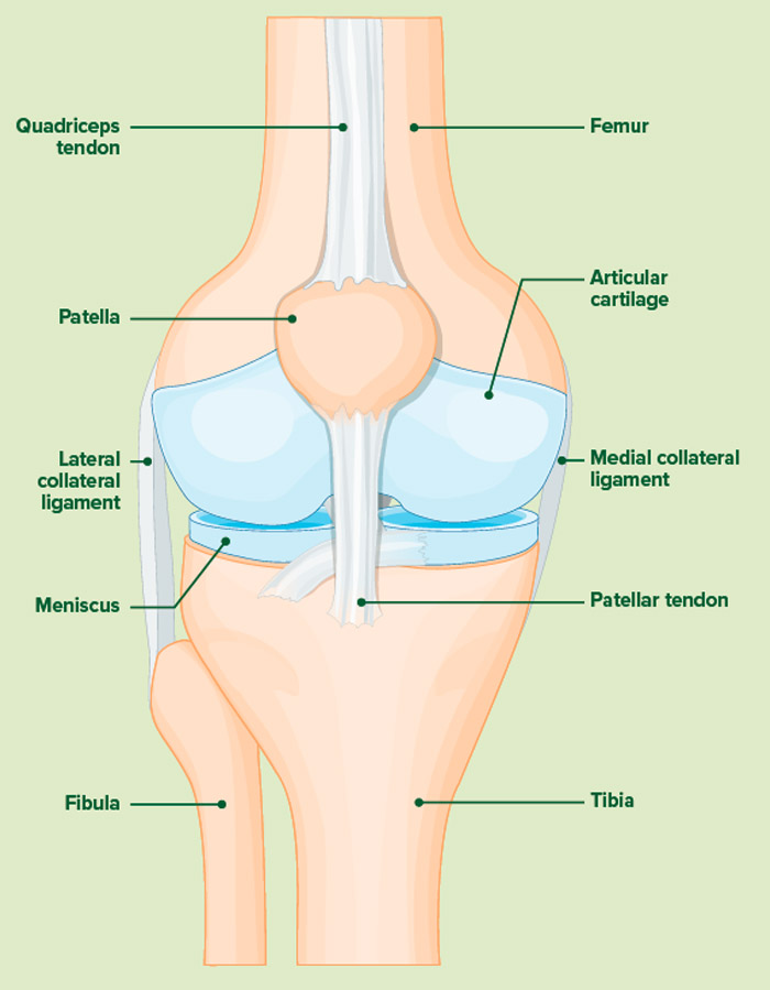
Joint movement is facilitated by cartilage, a smooth connective tissue that covers the ends of the bones. Some joints – those that allow the widest range of movement, such as the knees (as pictured) and wrists – also contain synovial fluid which provides extra lubrication and acts as a shock absorber.
Bones move when the attached muscles contract. Skeletal muscles can only pull in one direction, so usually exist in pairs. For example, contracting the bicep muscle in the upper arm allows the lower arm to flex towards the upper arm, and contracting the opposing tricep muscle allows it to move away again.
Tendons are tough bands of connective tissue that attach muscles to bones, while ligaments, which are similar in structure, hold bones together – in some cases restricting movement in order to protect a joint.