




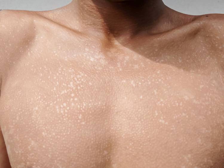
 17 Min
 In Clinical reviews

Follow this topic

Bookmark

Record learning outcomes
|
||||||||||||||
Superficial fungal infections are caused by dermatophytes and non-dermatophytes. They are among the most prevalent dermatological conditions, primarily affecting the skin, hair and nails.
One recent analysis estimated that there were approximately 1.73 billion cases of fungal skin infections worldwide in 2021. This article covers common fungal infections affecting the trunk, face and scalp; how pharmacists should assess patients; common differentials; and appropriate management.
Broadly, these infections are caused by two main organism groups: dermatophytes and Malassezia yeasts.
Dermatophyte infections, commonly known as tinea, are caused by highly infectious fungi that affect superficial keratinised tissues. These fungi require keratin to grow and release keratinase enzymes that digest it, so infection is normally limited to the epidermis.
Dermatophytes thrive when the skin is damaged by trauma, irritated or macerated, and when barrier function is compromised. The causative organisms include Trichophyton rubrum (most commonly), Trichophyton interdigitale and Trichophyton mentagrophytes. Clinical classification depends on the site of infection; for example, tinea affecting the body is termed tinea corporis.
|
||||||||||||
Tinea corporis
Patient assessment
A structured assessment for suspected tinea corporis should include questions about:
- Itching or localised burning
- Exposure to hot or humid environments, tight or occlusive clothing, obesity or hyperhidrosis
- Conditions that increase infection risk or complications such as diabetes or immunosuppression
- Similar symptoms elsewhere, such as the feet, nails or groin, which may be the primary source
- Similar symptoms in close contacts or contact with anyone known to be infected.
Clinical presentation
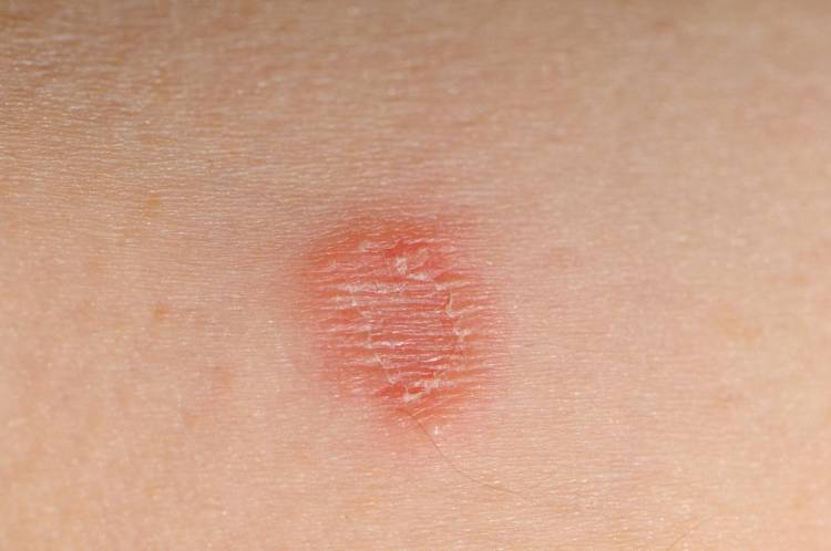
Tinea corporis classically affects non-hairy areas of the body, such as the trunk, neck, arms and legs. The fungus spreads laterally, presenting as round, pruritic, scaly plaques with central clearing and an active, inflamed, and sometimes raised border. This ring-like appearance gives rise to the common name ringworm.
| Tinea corporis lesions are often asymmetric but can be bilateral |
Lesions are often asymmetric but can be bilateral. It is helpful to ask whether patients have similar symptoms elsewhere, such as the nails, feet or groin. Diagnosis is usually straightforward, but common differentials and distinguishing features are shown in Table 1.
First-line treatment
Topical imidazoles:
- Clotrimazole: applied twice daily for up to 4 weeks
- Miconazole: applied twice daily and continued for 10 days after skin lesions have healed.
There is little evidence of meaningful differences among the available imidazoles.
Allylamines:
- Terbinafine: applied thinly to the affected area once or twice daily for 1 to 2 weeks.
Imidazoles are fungistatic and prevent further growth of the fungus. In contrast, allylamines are fungicidal and toxic to fungi. This difference in action explains why imidazoles typically require a longer treatment period. Antifungal therapy should be continued for 1 to 2 weeks after clinical symptoms have resolved to prevent reinfection.
Second-line treatment
Where infections are widespread or if patients are immunocompromised, oral therapy is required e.g. terbinafine 250mg daily for 4 weeks. The drug should be avoided in patients with either hepatic or renal impairment. Although oral therapy could be initiated by community pharmacist prescribers, GP referral is preferred to assess hepatic and renal function.
Self-care measures
As dermatophyte infections thrive in warm, moist environments, patients should be advised to wear loose-fitting clothing made from moisture-wicking materials. They should also avoid scratching lesions, as this may spread infection, and avoid sharing towels because of the risk of fungal transmission.
| Table 1: Common differentials for tinea corporis | |||
| Skin condition | Lesion border | Central clearing | Distinguishing features |
| Discoid eczema | Sharp, coin-like | Absent | Highly pruritic, present on extremities/symmetrical. Peak incidence age 50-60 years |
| Pityriasis rosea | Multiple, circular lesions affecting trunk and limbs, often symmetrical | Absent | Often preceded by a single, larger lesion (herald patch) about 1-2 weeks before other lesions develop |
| Granuloma annulare | Single or multiple rings of small, smooth, red or flesh-coloured papules | Present | No scale, often present on the dorsal surface of the hands or feet |
| Psoriasis | Inflamed plaques covered by adherent silvery-white scale | Absent | Plaques often symmetrical and present on extensor surfaces, scalp and associated with nail changes |
Pityriasis versicolor
Pityriasis versicolor (PV) is a harmless infection of the trunk caused by the commensal lipophilic yeast Malassezia. Colonisation with Malassezia increases with age, so that around 25% of children and almost 100% of adults are affected.
Although it is normally part of the skin flora, Malassezia can become pathogenic. PV is commonly seen in hot, humid climates and generally occurs in teenagers and young adults, with higher incidence during warmer weather.
Patient assessment
- Ask about pruritus, although it is mild or even absent in most cases
- Enquire about potential predisposing factors, including hyperhidrosis, use of occlusive clothing, dressings, lipid-rich emollients, malnutrition and immunosuppression or deficiency.
Clinical presentation
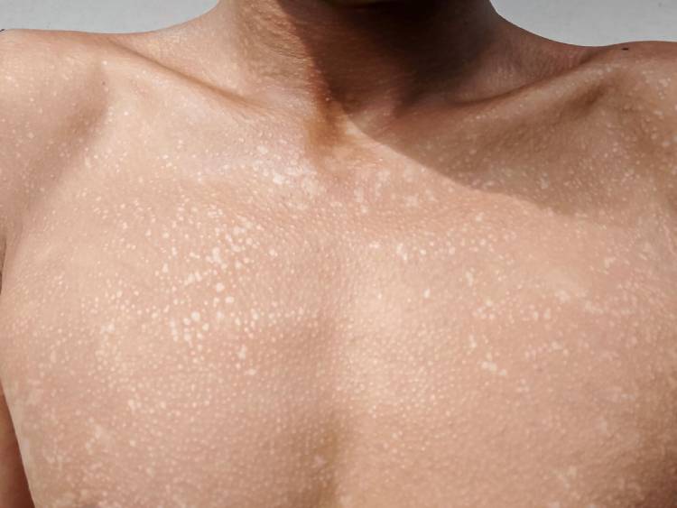
Patients often first notice PV after sun exposure, when oval-to-round hypopigmented macules become more apparent, particularly in people with skin of colour.
Lesions typically affect sebum-rich areas such as the neck, upper arms and abdomen, and sometimes the face; PV can also occur in the groin and axillae. In lighter skin tones, macules tend to be coppery brown with a small amount of scale.
Diagnosis is usually clinical but can be confirmed with a Wood’s lamp, which shows yellow-green fluorescence in affected areas. Common differentials are shown in Table 2. Although considered an infection, pityriasis versicolor is not transmissible.
Pityriasis versicolor may be mistaken for psoriasis, particularly guttate psoriasis, which presents with small, scaly macules across the trunk and limbs, or tinea corporis.
First-line treatment
For small areas, a topical imidazole applied twice daily for 2 to 3 weeks is effective. In more widespread cases, an antifungal shampoo such as ketoconazole 2% or selenium sulphide 2.5% can be used. The shampoo should be lathered, applied to the affected areas and left in contact with the skin for about 15 minutes before being washed off. This process should be repeated for five days.
Second-line treatment
Where topical therapy fails or is more difficult to apply, oral therapy can be tried. Choices include:
- Itraconazole 200mg once daily for 7 days
- Fluconazole 50mg once daily for 2 to 4 weeks, or 300-400mg weekly for 1 to 3 weeks.
Itraconazole should be avoided in those with liver failure or heart failure and in women who are pregnant or breastfeeding. Patients should be informed that it can take several months for the skin colour to return to normal, although occasionally the discolouration can be very persistent.
Self-care measures
Recurrences of pityriasis versicolor are common. For patients prone to recurrence, ketoconazole used for 5 to 10 minutes, one to four times a month, may help prevent further episodes. If symptoms recur after exposure to warm or humid environments, 2% ketoconazole shampoo can be applied once daily for up to three days before sun exposure.
| Table 2: Common differentials for pityriasis versicolor | |
| Skin condition | Distinguishing features |
| Vitiligo | Complete loss of pigmentation rather than hypopigmentation; no fluorescence under a Wood’s lamp |
| Pityriasis rosea | Often preceded by a single, larger lesion (herald patch) about 1-2 weeks before other lesions develop. Macules have slight scaling around the edges |
| Erythrasma | Tends to present as erythematous or hyperpigmented regions in the axillae or groin; coral-red under Wood’s lamp |
| Pityriasis alba | Normally ill-defined, pink patches with slight scale, commonly seen on the face in children and young adults with atopic eczema |
Malassezia folliculitis
Malassezia folliculitis (MF) is an inflammatory acneiform eruption of the pilosebaceous unit. The condition is common in adolescents, probably because of increased sebaceous gland activity, and is caused by the same yeast responsible for pityriasis versicolor. It often affects people living in hot, humid climates who sweat frequently.
Patient assessment
- Ask about pruritus, as most patients will report intense, persistent itching
- Also ask about predisposing risk factors, such as sweat-inducing activities (e.g. gym sessions), occlusive clothing, hot and humid working environments (e.g. kitchens), and immunosuppression
- Enquire about recent use of topical or oral broad-spectrum antibiotics for acne, or topical and oral corticosteroids, which are recognised risk factors.
Clinical presentation
Malassezia folliculitis is characterised by intensely pruritic, 1-2mm, monomorphic, follicular, dome-shaped papules and pustules, predominantly affecting seborrhoeic areas such as the upper back, chest, face and shoulders. The main differentials are shown in Table 3.
MF is often mistaken for acne and can develop after inappropriate topical or oral acne therapy with broad-spectrum antibiotics, because these treatments reduce bacterial flora and allow the yeast to multiply unchecked.
First-line treatment
Mild cases respond well to topical imidazoles such as ketoconazole 2% shampoo, applied three to five times a week (left in contact with the skin for 10 minutes) until cleared.
Second-line treatment
Oral antifungals include:
- Fluconazole: 100-200mg orally once daily for 14-21 days, or
- Itraconazole: 100mg orally twice daily for 14 days.
Self-care measures
All patients with MF should avoid lipid-rich emollients and greasy skincare products because these lipids act as a food source for the yeast. Showering after intense exercise is also advisable, as humidity and occlusion can trigger MF. Breathable fabrics may reduce heat and sweating in sebaceous areas such as the chest and back, which are common sites of infection.
| Table 3: Common differentials for Malassezia folliculitis | |
| Skin condition | Distinguishing features |
| Acne | Presence of both open and closed comedones that are absent in malassezia folliculitis. Additionally, acne lesions are rarely pruritic |
| Steroid-induced acne | Though monomorphic, lesions tend to be sore and much less pruritic. Can present with nodules or cysts |
| Bacterial folliculitis | Though lesions are monomorphic, these tend to be either tender or painful rather than pruritic, and occur mainly in areas of hair growth |
Seborrhoeic dermatitis
Seborrhoeic dermatitis (SD) is a chronic inflammatory skin disorder affecting areas of the body with a high concentration of sebaceous glands including the scalp and face. While the precise cause is unclear, it is associated with Malassezia yeasts. It occurs in infants, mainly on the scalp, where it is known as cradle cap.
Patient assessment
A structured assessment for suspected seborrhoeic dermatitis should include questions about:
- Scalp involvement, including pruritus and scaling
- Known triggers, e.g. environmental (both cold and high humidity), emotional and infectious
- Onset, which is normally after puberty, and whether the disease follows the typical relapsing-remitting pattern
- Use of medicines that can trigger the condition, such as cimetidine, gold and methyldopa, or specific illnesses in older adults, e.g. Parkinson’s disease and HIV.
Clinical presentation
Seborrhoeic dermatitis is characterised by erythematous, greasy, scaly patches on the scalp, nasolabial folds, ears, eyebrows, eyelids and chest. Other affected areas include the postauricular area, forehead and anterior chest. Common differentials for seborrhoeic dermatitis are shown in Table 4.
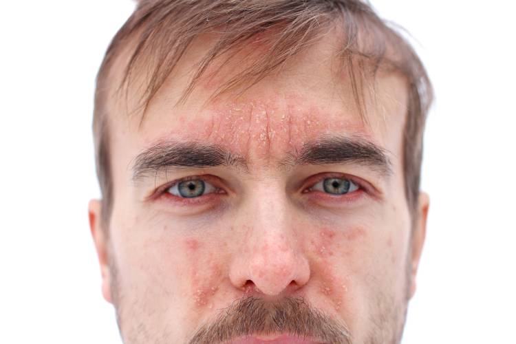
| The exact cause of seborrhoeic dermatitis is unclear — but it is associated with Malassezia yeasts |
First-line treatment
For scalp involvement in infants, barrier emollients such as zinc and castor oil ointment can be effective. In children and adults, shampoos containing salicylic acid, ketoconazole, selenium sulphide, coal tar or zinc pyrithione are effective.
For non-scalp areas, topical imidazoles such as ketoconazole (applied twice daily) for up to 4 weeks are effective. Adding a short course of a low-potency topical steroid, e.g. hydrocortisone 1%, can help reduce inflammation and pruritus.
Second-line treatment
For more widespread exposure, oral antifungals are effective, e.g. itraconazole: 100-400mg/day orally, given in two divided doses. Oral ketoconazole should be avoided due to concerns of hepatotoxicity.
Self-care measures
Given the chronic and relapsing-remitting nature of seborrhoeic dermatitis, self-care measures are designed to manage acute disease flares.
Ketoconazole shampoo can be used as a face and scalp wash. Using light emollients and avoiding lipid-rich products, which feed Malassezia yeasts, can also help.
Scalp involvement
Seborrhoeic dermatitis may also affect the scalp. In its mildest form, it causes dandruff with diffuse, fine white scaling. As it progresses, poorly demarcated erythematous plaques with greasy, yellowish scale may develop on the anterior hairline and retroauricular folds. Treatment is with shampoos as described above.
| Table 4: Common differential diagnoses for seborrhoeic dermatitis | |
| Skin condition | Distinguishing features |
| Rosacea | Presents with central facial erythema. Lacks the greasy yellow scaling characteristic of SD and has specific triggers (e.g. alcohol) absent for SD |
| Psoriasis | Often presents on face with drier, thicker, silvery-white scales alongside plaques on elbows, knees or scalp. Lacks greasy, yellowish scale seen in SD |
| Tinea faciei (facial tinea) | Fine, white scale, often asymmetric (SD is symmetric) and annular with active border |
| Atopic dermatitis | Often present on creases (e.g. eyelids; around the mouth). Skin is very dry and intensely pruritic. Comorbidities include asthma/hay fever |
Tinea capitis
This is predominantly seen in children and young teens aged 3 to 14 years. The fungi can penetrate the hair follicle’s outer root sheath and ultimately may invade the hair shaft.
Patient assessment
- Ask about hair loss, scaling and pruritus, which are common features
- Also enquire about inflammation and pustule formation, which may also occur
- Examine the scalp for any black dots caused by broken-off hairs.
Clinical presentation
Its appearance varies. Non-inflammatory disease causes diffuse scaling and pruritus, with single or multiple patches of asymmetric alopecia and a black-dot appearance.
By contrast, inflammatory disease can cause erythema, numerous pustules and crusting, sometimes with a painful, boggy, pus-filled kerion several centimetres in diameter and localised alopecia. Some individuals may be systemically unwell. Common differentials for tinea capitis are shown in Table 5. Tinea capitis is infectious and can spread to others.
| Table 5: Common differentials for tinea capitis | |
| Skin condition | Distinguishing features |
| Seborrhoeic dermatitis | No evidence of hair loss, surface scale tends to be greasy. Non-inflammatory tinea capitis presents with dry, powdery scale |
| Scalp psoriasis | Psoriatic plaques are well demarcated and frequently extend beyond the anterior hairline onto the forehead. Scale is dry and silvery-white. Scraping the scale reveals small bleeding points. Hair growth is normal |
| Alopecia areata | The skin within the areas of hair loss feels perfectly smooth, normal, and lacks erythema, scaling or pustules |
| Atopic eczema | Often causes a scaling with pruritus. Hair loss is less common and there is often a personal or family history of atopy |
Management
Oral antifungal agents are required, as topical therapy cannot penetrate the hair shaft:
- Adults: griseofulvin 1,000mg once a day or 500mg twice a day for 4-8 weeks
- Children 12-17 years: griseofulvin 500mg daily, increased if necessary to 1g daily for severe infections
- Children 11 months to 11 years: griseofulvin 10mg/kg daily (maximum per dose 500mg), increased if necessary to 20mg/kg daily (maximum per dose 1g) for severe infections.
Doses in children can be reduced once a clinical response has occurred.
In addition to oral therapy, the likelihood of tinea capitis spreading can be reduced with topical therapy using selenium sulphide shampoo applied twice weekly for two weeks, leaving it in contact with the scalp for 2 to 3 minutes. Alternatively, ketoconazole shampoo can be used 2 to 3 times weekly for 2 to 4 weeks, again leaving it in contact with the scalp for a few minutes.
Self-care measures
Surface crusts can be softened and gently removed. Potential sources of fungal spores, such as hats, scarves, hairbrushes and combs, should be discarded or disinfected to prevent reinfection. Towels should not be shared and parents should inspect children’s scalps for signs of infection. If pets are suspected as the source, they should be assessed and treated by a vet.
References & further reading
- Li D, Fan S, Zhao H, et al. Worldwide trends and future projections of fungal skin disease burden: a comprehensive analysis from the Global Burden of Diseases study 2021. Front Public Health. 2025 Jun 4; 13:1580221
- Leung AK, Barankin B, Lam JM, Leong KF, Hon KL. Tinea pedis: an updated review. Drugs Context. 2023;12:2023-5
- Elewski BE, Haley HR, Robbins CM. The use of 40% urea cream in the treatment of moccasin tinea pedis. Cutis. 2004;73(5):355-7
- Faergemann J, Baran R. Epidemiology, clinical presentation and diagnosis of onychomycosis. Br J Dermatol. 2003;149 Suppl 65:1-4
- Leung AKC. Pityriasis versicolor. In: The Encyclopedia of Molecular Mechanisms of Disease (Lang F, editor). Berlin: Springer-Verlag; 2009. pp. 1652-1654
- Durdu M, Güran M, Ilkit M. Epidemiological characteristics of Malassezia folliculitis and use of the May-Grünwald-Giemsa stain to diagnose the infection. Diagn Microbiol Infect Dis. 2013;76(4):450-7

Share:


In Clinical reviews